
The patient guide · Breast
Breast Implant Options in NYC
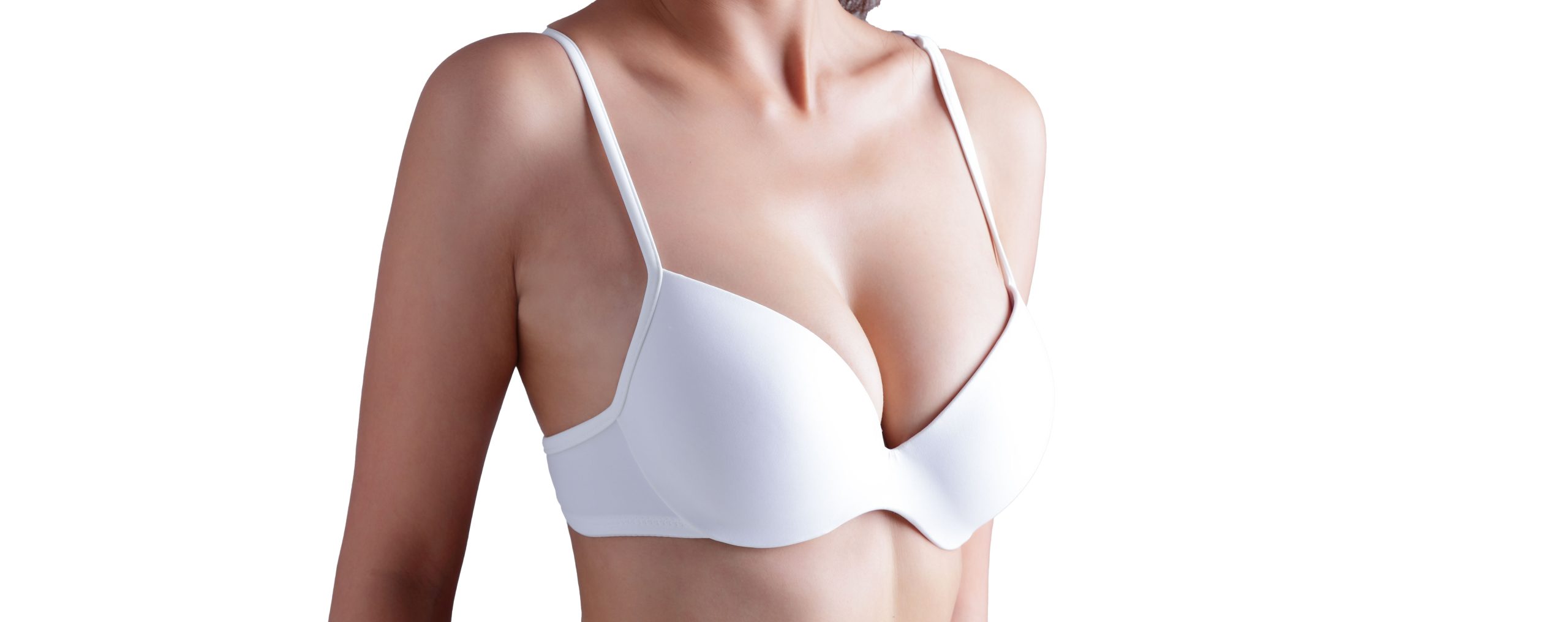
A considered guide to your options, the questions that matter, and the conversation ahead.
In this guide
What you are actually choosing
Watch one patient describe her size goals and consultation experience.
Breast augmentation is not one decision. It is a set of them, and they interact: what the implant is filled with, how large it is, what shape it is, how far it projects, what its surface is like, whether it sits over or under the chest muscle, and where the incision is placed.
Which combination is right depends on measurements rather than preference alone. The width of your chest and of your breast base, how much breast tissue you have and where it sits, the quality and thickness of your skin, the position of the fold beneath the breast, and any existing asymmetry all constrain what will work. Dr. Smith takes those measurements at consultation and works through the options with you, with reasons, rather than handing down a single recommendation.
Seeing the options before you decide
The practice uses the Canfield Vectra system for three-dimensional imaging. Photographs are captured and rendered as a three-dimensional model of your own anatomy, and different implant sizes, shapes and profiles can be simulated on it. It is a planning tool rather than a promise of a result, and it is most useful for narrowing a range and for showing what a particular volume actually does on your frame, which is difficult to judge from photographs of other people.
Where the choices constrain each other
Some of these decisions limit others. A narrow chest limits how much volume can be added before the implant sits wider than the breast that covers it. Thin tissue at the upper pole limits how well the edge of an implant can be concealed, which affects both the fill material and the placement. A preference for a particular look sometimes points to a different implant than the one a patient arrived expecting. Dr. Smith will say plainly where that is the case.
Silicone or saline
Both are available, and silicone is what Dr. Smith uses in the vast majority of cases.
Silicone implants
A silicone implant is a silicone shell filled with silicone gel. It is the more common choice because the gel more closely resembles breast tissue in feel, and because visible rippling at the edges is less likely.
What are gummy bear breast implants?
“Gummy bear” is a nickname for form-stable, highly cohesive silicone gel implants. They are silicone implants, not a third fill option alongside silicone and saline. Cohesivity describes how strongly the gel holds together. It does not tell you the implant’s shape, shell surface, profile or brand. Cohesive gel is used in round as well as shaped designs.
More cohesive gel generally holds its form more firmly, but the firmest implant is not automatically the best match. How an implant feels and looks also depends on the complete device design and the breast tissue covering it. Compare the specific implants being considered, rather than choosing by the nickname alone.
| Decision | What to compare |
|---|---|
| Fill and gel | Saline or silicone gel; within silicone, ask how the proposed gel’s cohesivity affects feel and shape retention. |
| Shape | Round or anatomic describes the device’s shape. A cohesive gel does not automatically mean a teardrop-shaped implant. |
| Shell surface | Smooth and textured describe the outside of the implant, separately from the gel inside. Surface has its own risk considerations. |
| Profile and volume | Profile describes forward projection; volume describes size. Compare these with implant width and your breast measurements. |
A silicone implant rupture may not cause an obvious change in how the breast looks or feels. Many ruptures are silent, which is why follow-up imaging is recommended even without symptoms. A cohesive gel does not make an implant rupture-proof or remove the need for monitoring. Discuss implant risks, including rupture, capsular contracture, infection and possible further surgery, before deciding.
Saline implants
A saline implant is a silicone shell filled with sterile salt water after the shell has been placed, which allows a smaller incision. If a saline implant ruptures, fluid can leak out quickly or over several days, causing the implant to lose size or shape. A suspected rupture should be evaluated by your surgeon. The trade-off is feel: saline implants are firmer than silicone and show rippling more readily, particularly in patients with little breast tissue covering them.
Manufacturers
Ask which manufacturer and exact implant model are being proposed, and why that device fits your measurements and goals. Review its current patient labeling, including the boxed warning and patient decision checklist. A brand name or the term “gummy bear” is not enough to identify the implant’s design, risks or follow-up requirements.
How long implants last
Breast implants are not lifetime devices. How long an individual implant will remain in place cannot be predicted, and the chance of complications increases with time. Removal or replacement may be needed because of a complication or a change in your goals. Plan for ongoing follow-up and the possibility of future surgery; a “gummy bear” label does not establish a lifespan or replacement date.
Imaging after silicone implants
For people with silicone implants who have no symptoms, FDA guidance recommends the first ultrasound or MRI five to six years after placement, followed by screening every two to three years. New swelling, pain, a lump or a change in breast shape should be evaluated without waiting for that screening interval. Implant-rupture screening does not replace routine breast cancer screening. Saline implants do not require this routine screening for silent rupture.
Shape, profile and surface
Round or anatomic
Round describes the implant’s shape; it does not guarantee equal fullness in the upper and lower breast. The result also depends on the implant’s dimensions, gel, placement and your own tissue. Dr. Smith uses round implants for new breast augmentation.
Anatomic implants, also called shaped implants, have a teardrop shape with more volume in the lower portion. They are described here to explain the terminology, not as an option Dr. Smith offers for new breast augmentation. Cohesive silicone gel does not require an anatomic shape.
Profile
Profile describes how far an implant projects forward from the chest wall for a given volume. Manufacturers use their own terms, but the general categories are:
- Low profile. The least projection for a given volume, spread over the widest base.
- Moderate profile. Intermediate between the two. The majority of the implants Dr. Smith uses fall in this range.
- High profile. The most projection for a given volume, on the narrowest base.
Which profile suits you follows mainly from the width of your breast base. A low profile implant tends to read as less operated. A high profile implant is useful in a narrow breast, because it allows volume to be added forward rather than sideways, where it would sit wider than the breast covering it.
Smooth or textured
Dr. Smith uses only smooth, round implants for new breast augmentation, including Motiva SmoothSilk implants. Motiva classifies its SmoothSilk surface as smooth under the ISO surface standard; the “nano” terminology used for its surface design does not mean a conventional textured implant.
Textured shells were designed to adhere to the surrounding tissue and help hold a shaped implant in position. Capsular contracture, or tightening of the scar tissue around an implant, remains a possible complication with either implant surface.
Breast implant-associated anaplastic large cell lymphoma (BIA-ALCL) is an uncommon cancer of the immune system. FDA guidance identifies textured implants as having greater BIA-ALCL risk than smooth implants. The FDA has also received reports involving smooth implants, but previous implant exposure is often incompletely documented. Reports alone cannot establish which device caused the disease. Discuss the risks of each implant surface with Dr. Smith when choosing an implant.
In the three-year core-study results presented in Motiva’s 2024 FDA-reviewed labeling, no BIA-ALCL cases were reported. That finding does not establish zero long-term risk. The labeling retains a BIA-ALCL warning, and the risks remain part of the implant discussion.
Where the implant sits
Watch Dr. Smith explain why upper-body training matters when discussing implant placement.
The implant can go under the breast tissue and over the chest muscle, or under the pectoralis muscle. Under the muscle is what is used in most cases.
Submuscular placement
Placing the implant under, or partly under, the pectoralis major puts more tissue between the implant and the skin. That gives a softer transition at the upper edge, makes visible rippling less likely, is associated with a lower rate of capsular contracture, and makes mammograms easier to interpret.
The early muscle soreness can differ between implant positions, but Dr. Smith uses the same exercise milestones for conventional augmentation under or over the muscle when no lift is performed: light lower-body and core exercise at two weeks, light upper-body exercise at four weeks, and a gradual return to full exercise, including heavy lifting and running, from six weeks with clearance. The planner uses these exercise milestones when a breast lift is included as well; recovery still depends on the complete operation and individual healing. His breast augmentation recovery guide explains the full schedules for conventional augmentation, Preservé and breast lift.
Subglandular placement
Placing the implant over the muscle gives more direct access if further surgery becomes necessary. The implant is easier to see and to feel through the skin, particularly in a patient with little breast tissue, and the rate of capsular contracture is higher.
The situation in which over the muscle is clearly the better answer is a patient with a very developed pectoralis muscle, such as a competitive athlete or a bodybuilder. In those patients, flexing a strong pectoralis distorts an implant placed beneath it, which is called animation deformity. Outside that group it is unusual for subglandular placement to be the better choice.
Where the incision goes
An incision is needed to place an implant, and it will leave a scar. There are three usual locations, and they differ in how well the scar hides and in how much control the surgeon has over the pocket the implant sits in.
In the breast fold
The inframammary approach places the incision in the fold beneath the breast. It gives direct access to the pocket and the most control over exactly where the implant sits, and the scar is covered by the breast itself. This is the approach used most often.
At the border of the areola
The periareolar approach places the incision where the pigmented skin of the areola meets normal breast skin, usually along the lower half of that border. In a suitable patient the scar camouflages extremely well at the color transition. It carries a higher rate of capsular contracture and of infection than the inframammary approach, and it is not well suited to a small areola.
In the underarm
The transaxillary approach places the incision in the underarm, so there is no scar on the breast at all. It gives less direct control over the implant pocket and carries a somewhat higher rate of infection and capsular contracture than the inframammary approach.
How scars behave
Scars are raised and pink at first and fade substantially over the first year, after which they are generally difficult to see. How any individual scar matures varies between patients and cannot be predicted precisely beforehand, which is worth knowing before choosing an approach on the basis of the scar alone.
Questions to bring to your implant consultation
- Which exact implant model are you proposing, and why does it fit my breast width and tissue coverage?
- What would change if we chose a softer or firmer gel, a different profile or a different volume?
- What are the device-specific risks, and how do the shell surface and surgical plan affect the discussion?
- What follow-up imaging and possible future surgery should I plan and budget for?
For the financial side of the decision, see breast implant costs in NYC. Compare the complete surgical quote and ask separately about future imaging and revision costs; the implant nickname alone does not tell you what your care will cost.
Every one of the decisions on this page is made together with Dr. Smith at consultation, on the basis of your measurements, your tissue and what you want. The consultation fee is $500 and is applied toward any procedure you go on to book.
Related procedures
For information about Dr. Smith’s approach with Motiva implants, explore Preservé breast augmentation in NYC.
Written by Darren M. Smith, MD, FACS, board-certified plastic surgeon, Manhattan.
YOUR NEXT CHAPTER
Care begins
with listening.
Discuss your priorities, ask questions and explore treatment options with our team.
Our practice is located at the Ritz Tower, 111 East 57th Street in Midtown Manhattan. We welcome patients from the five boroughs and the surrounding tri-state area. If you are traveling for care, explore our guide for out-of-town patients.
Request a consultation